Advances in Atomic Force Microscopy: PinPoint Piezo Force Microscope - frictionless imaging technique

Advances in Atomic Force Microscopy:
PinPoint Piezo Force Microscope – frictionless imaging technique
(Seminar and Life Demo)
When:
- Tuesday, 20 November, 14.30-16.30, Room Boston
Materiaux, 19-23 November 2018, Strasbourg / France

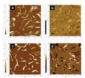
Conventional PFM(a,b) vs. PinPoint PFM(c,d)

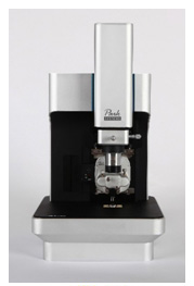
NX10 AFM
Program
- 14:30 Welcome and Introduction
- 14:40 Talk: “PinPoint Piezo Force Microscope – frictionless imaging technique “
- 15:30 Instrument Demonstration on Park NX10 AFM
- 16:15 Discussion und Summary
ABSTRACT
Electromechanical coupling in materials is a key property that provides functionality to a variety of applications including: sensors, actuators, IR detectors, energy harvesting, and biology. Most materials exhibit electromechanical coupling in nanometer-sized domains. Therefore, to understand the relationships between structure and function of these materials, characterization on the nanoscale is required. This electromechanical coupling property can be directly measured in a non-destructive manner using piezoelectric force microscopy (PFM), a well – established method in atomic force microscopy (AFM).
The conventional PFM is usually performed in contact mode. Contact mode, however, has challenges for a great variety of samples, e.g. the characterization of an annealed phenanthrene thin film on top of an ITO surface. The main difficulty in imaging these samples is due to the phenanthrene thin film forming rod-shaped nanostructures that are very susceptible to displacement by a scanning AFM probe. This is the reason for the newly developed PinPoint PFM mode by Park Systems. As opposed to standard contact mode, in PinPoint mode the AFM probe monitors its feedback signal, approaches towards the sample surface until a predefined force threshold is reached, and measures the Z scanner’s height. The AFM probe is then rapidly retracted away from the surface to a user-defined height. During the data acquisition the XY scanner stops movement completely and therefore no AFM probe movement takes place on the sample surface at any time. This allows for a more accurate representation of the surface as the nanorods are not moved from their original position.
Join the seminar and live demo to learn more about advantages of PinPoint PFM over a conventional PFM mode.
SPEAKER INFORMATION:
Victor Bergmann – Application Scientist at Park Systems
pse@parksystems.com | www.parksystems.com
